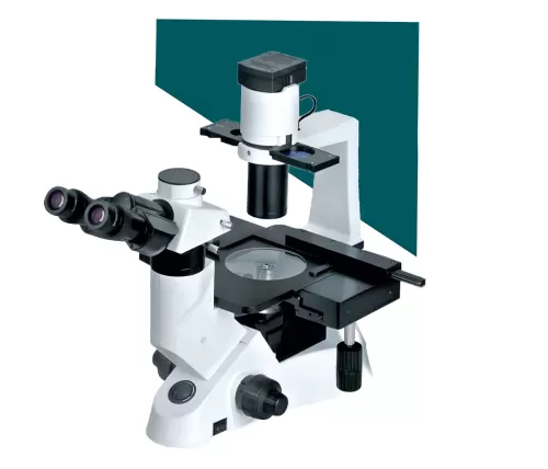
INVERTED FLUORESCENCE MICROSCOPE (TM-10)
PC Configuration: Core i5 processor, 13th generation, RAM 16GB, 2TB HDD, 2GB Media Graphic Card, Display resolution 1920x1024, 32-bit, drive DVD-ROM drive, TFT LED screen 23-inch monitor Multimedia kit, OS, Windows 10
INVERTED FLUORESCENCE MICROSCOPE (TM-10)
Microscope Body:
Inverted trinocular microscope with Infinity corrected optical system with light distribution between eyepiece and camera port of 100:0/ 0:100
Observation technique Brightfield, Phase contrast & Fluorescence. Microscope can be ungradable to DIC, Emboss Contrast(pseudo3-D) observation technology.
Eyepiece:
10X with FOV23 mm and diopter adjustment facilities on both eyes, anti-fungus type
Condenser:
Focusable ELWD N.A-1.25, Focusable,
Phase slider centerable, brightfield, fluorescence, Phase centering telescope or better
Nosepiece: Encoded 4 position, Quadruple nosepiece, DIC slot
Stage:
Attachable mechanical stage with universal holder to accepting all types of specimen holders.
Illumination:
High intensity uniform brightness distribution (20-30W power consumption)
cool white LED light, with time of >60,000hrs.
Objectives:
Long working distance objectives with specialized phase contrast, Brightfield and Fluorescence Imagining.
• Plan Achromat 4X, N.A 0.10, W.D.30.mm.
• Plan Achromat Phase10x, NA0.25, Ph1 FWD 3-4 mm
• Plan Achromat 20X NA0.30, Ph1 FWD 3.5-4.5 mm
• Plan flour LWD 40X, NA 0.40, FWD 2.0-2.2 mm
• System can be upgraded to 100X oil magnification as research required.
Fluorescent attachment:
Epi-fluorescence rotating filter turret (with main body), Filter cubes with noise terminator mechanism, configured with up to 3 Epi-fluorescence filter cubes, Additional positions for bright-field observation,
Fluorescence illumination of LED Filter set of DAPI, FITC and TRITC.LED lights source of over 10,000 hour’s life time. Fluorescence Filters and Fluorescence LED can be synchronized while changing the filter to reduce
photo bleaching. Automatic LED fluorescence illumination intensity recognition function. Special feature to replicate illumination power of same wavelength to reduce minimum adjustment.
Camera:
Digital CMOS Camera system: Scientific microscopic digital CMOS color Camera system: High
resolution scientific CMOS camera of sensor size (1/1.8 inch & image pixel size 2.0x2.0 microns) QE 60%, resolution of 5.9 MP, 10-15frame per-second live display, Live cell imaging, USB 3.0 Pc interface, Exposure time:100usec to 30sec, Exposure control: One-push auto exposure.0 mount is provided.
Camera can capture Fluorescence images.
Software:
Imaging software for PC imaging, Fluorescence channel mixing/un-mixing, image analysis, live cell imaging bar insert, annotation, shading correction, image overlay for fluorescence & intensity measurement, automatic counting, manual counting, image enhancement through brightness, contrast, smooth/sharpen, background subtraction, white balance
live measurement such as length, width, area, perimeter, etc.
PC Configuration: Core i5 processor, 13th generation, RAM 16GB, 2TB HDD, 2GB Media Graphic Card, Display resolution 1920x1024, 32-bit, drive DVD-ROM drive, TFT LED screen 23-inch monitor Multimedia kit, OS, Windows 10